Understanding Hip Labral Tears
Hip labral tears often stem from femoroacetabular impingement (FAI), impacting the ball-and-socket joint where the femoral head meets the acetabulum within the pelvis.
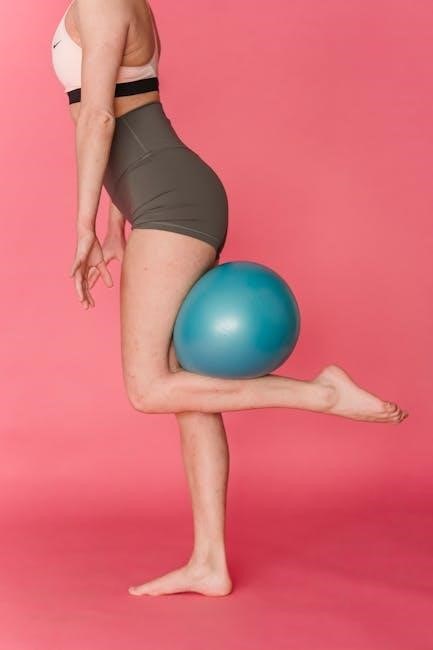
Initial nonoperative treatment, lasting around three months, focuses on core strengthening and aims to resolve the issue before considering surgical interventions.
What is the Hip Labrum?
The hip labrum is a crucial component of the hip joint, functioning as a fibrocartilaginous ring that deepens the acetabulum – the socket within the pelvis. This ring significantly enhances the stability of the hip, contributing to a secure fit between the femoral head (the ball at the top of the thigh bone) and the acetabulum.
Essentially, it acts like a bumper, preventing the femoral head from dislocating and distributing weight-bearing forces across the joint. The hip joint is formed by the convergence of the ilium, pubis, and ischium, creating this vital socket. Damage to this labrum, a hip labral tear, can lead to pain, instability, and limited range of motion, often requiring a comprehensive rehabilitation protocol. Understanding its anatomy is key to effective treatment.
Causes of Hip Labral Tears
Hip labral tears frequently arise from structural abnormalities, most notably femoroacetabular impingement (FAI). This occurs when there’s abnormal contact between the femoral head and the acetabulum during hip movement. Repetitive motions, common in athletes, can exacerbate this impingement, leading to labral damage over time.
Trauma, such as a sudden impact or twisting injury, can also directly cause a tear. The hip’s complex anatomy – involving the ilium, pubis, and ischium forming the acetabulum – makes it vulnerable. Additionally, hip dysplasia (a shallow socket) or even subtle anatomical variations can predispose individuals to labral tears. Recognizing these underlying causes is vital for targeted rehabilitation and preventing recurrence.
Symptoms of a Hip Labral Tear
Symptoms of a hip labral tear can vary significantly in presentation. A common complaint is groin pain, often described as a deep ache, which may radiate to the buttock or thigh. This pain is frequently aggravated by activities like walking, squatting, or twisting motions. Individuals might experience a clicking, locking, or catching sensation within the hip joint, indicating mechanical symptoms.
Pain can also be present with prolonged sitting. The hip, a weight-bearing joint formed by the femoral head and acetabulum, may feel unstable or give way. It’s important to note that symptoms can mimic other conditions, making accurate diagnosis crucial. Early recognition allows for prompt intervention and management of discomfort.
Non-Surgical Treatment Options
Nonoperative treatment, typically employed for three months, centers on core strengthening exercises and aims to alleviate pain and potentially resolve the labral tear.
Initial Conservative Management (3 Months)
Initial management for a suspected hip labral tear prioritizes a three-month period of conservative treatment before considering more invasive options. This phase focuses on reducing pain, inflammation, and improving hip joint mechanics through non-surgical interventions.
A core component involves a carefully designed exercise program emphasizing core stabilization. Strengthening the muscles surrounding the hip and pelvis provides support and improves control. This often includes exercises targeting the glutes, abdominals, and lower back.
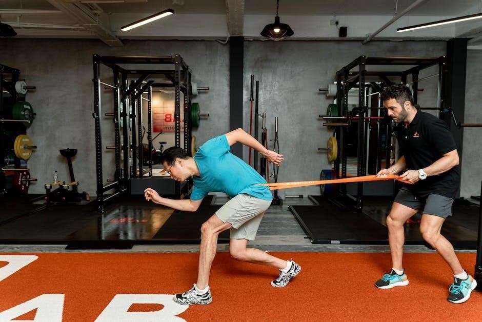
Activity modification is crucial; patients are advised to avoid activities that aggravate symptoms. Physical therapy plays a vital role, guiding patients through appropriate exercises and providing education on proper body mechanics. The goal is to potentially resolve the issue without surgery.
Pain Management Strategies
Effective pain management is paramount during the initial stages of hip labral tear treatment. Strategies often begin with over-the-counter pain relievers, such as NSAIDs (non-steroidal anti-inflammatory drugs), to reduce pain and inflammation.
Rest and activity modification are essential; avoiding aggravating activities allows the hip joint to recover. Ice application can help minimize pain and swelling, particularly after activity. In some cases, a corticosteroid injection may be considered to provide temporary pain relief and reduce inflammation, facilitating participation in physical therapy.
However, injections are typically not a long-term solution. Physical therapy focuses on restoring proper hip mechanics and strengthening supporting muscles, ultimately aiming to reduce pain and improve function.
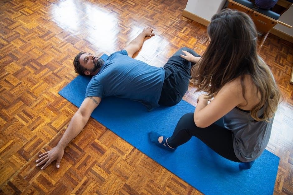
Phase 1: Early Rehabilitation (0-6 Weeks)
Initial rehabilitation prioritizes reducing pain and inflammation, focusing on gentle movements and isometric exercises to activate hip muscles without stressing the labrum.
Goals of Phase 1
The primary goals during the initial 0-6 week phase of rehabilitation following a hip labral tear, or after arthroscopic surgery for FAI, center around pain and swelling management. Reducing discomfort is paramount, allowing for gradual increases in activity. Restoring a pain-free range of motion is also crucial, beginning with gentle movements and progressing as tolerated.
Early phase exercises aim to activate the muscles surrounding the hip joint – glutes, hip flexors, and abductors – without placing undue stress on the repaired or irritated labrum. Isometric contractions are favored, building strength without joint movement. Neuromuscular control is initiated, focusing on regaining awareness of hip position and movement. Protecting the healing tissues is vital, avoiding provocative movements that could re-injury the labrum. Ultimately, Phase 1 establishes a foundation for more advanced rehabilitation stages.
Gentle Range of Motion Exercises
Initial range of motion (ROM) exercises in Phase 1 focus on restoring movement without exacerbating pain. Heel slides gently mobilize the hip through flexion and extension, performed lying down. Supine hip rotations, both internal and external, improve rotational mobility, keeping movements small and controlled. Abduction and adduction, performed while supine, enhance side-to-side movement.
Emphasis is placed on pain-free movement; any discomfort signals the need to reduce the range. Avoidance of deep hip flexion, adduction, or rotation is crucial to protect the labrum. Gentle hip circles can also be incorporated, promoting overall joint mobility. These exercises are typically performed multiple times daily, gradually increasing the repetitions as tolerated, always prioritizing controlled, pain-free motion.
Isometric Exercises for Hip Muscles
Isometric exercises are foundational in Phase 1, strengthening hip muscles without joint movement, minimizing stress on the labrum. Hip abduction isometrics involve squeezing a pillow between the knees, holding for 5-10 seconds. Adduction isometrics utilize similar resistance, focusing on inner thigh engagement. Gluteal squeezes activate the buttocks, improving hip extension stability.
Hip flexion isometrics can be performed by gently attempting to lift the leg while resisting the movement with a hand. These exercises should be pain-free and held without straining. Repetitions are typically performed in sets of 10-15, several times a day. Isometric holds build foundational strength, preparing the hip for more dynamic movements in subsequent rehabilitation phases.
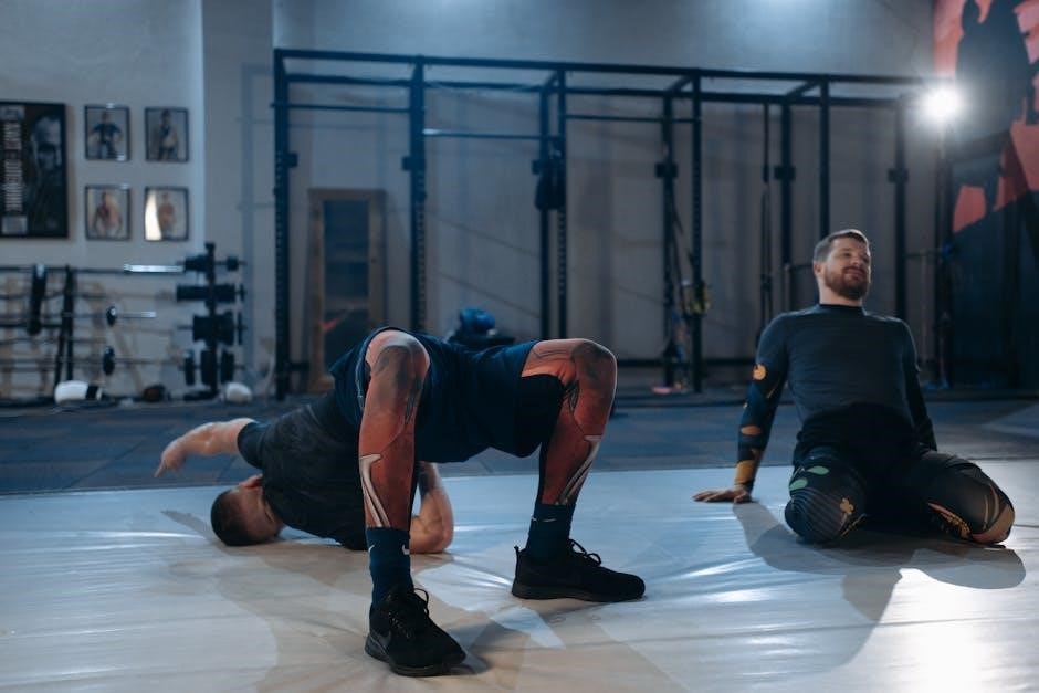
Phase 2: Intermediate Rehabilitation (6-12 Weeks)
Progressive strengthening, core stabilization, and proprioceptive training are key during this phase, building upon the foundation established in early rehabilitation.
Progressive Strengthening Exercises
Progressive strengthening in Phase 2 focuses on gradually increasing the load and complexity of exercises. Begin with hip abduction and adduction using resistance bands, progressing to weight machines as tolerated.
Bridging exercises, both single and double leg, are crucial for gluteal activation and core stability. Introduce clam shells with resistance bands to target hip external rotators.
Step-ups onto a low platform, focusing on controlled movement, enhance functional strength. Squats, initially bodyweight and then with added weight, improve overall lower extremity strength.
Remember to prioritize proper form and avoid any movements that provoke pain. The goal is to build strength without exacerbating the labral tear or causing further irritation. Listen to your body and adjust the intensity accordingly.
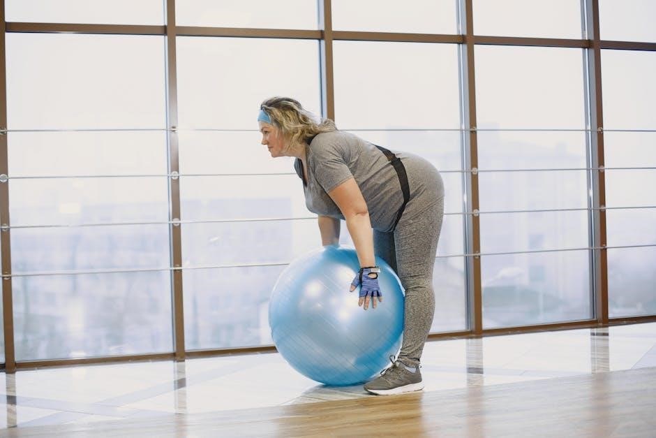
Core Stabilization Exercises
Core stabilization is paramount in Phase 2, providing a foundation for hip function and reducing stress on the labrum. Begin with pelvic tilts, focusing on engaging the transverse abdominis. Progress to dead bugs, maintaining a neutral spine while coordinating limb movements.
Bird dogs challenge core stability while promoting spinal alignment. Plank variations, starting with forearm planks and progressing to high planks, build endurance.
Side planks target the obliques, crucial for hip stability during lateral movements. Incorporate anti-rotation presses with resistance bands to resist rotational forces.
Focus on maintaining proper form and controlled breathing throughout each exercise. A strong core helps to control pelvic motion and optimize hip mechanics, supporting the healing process.
Proprioceptive Training
Proprioception, or body awareness, is vital in Phase 2 to restore neuromuscular control around the hip joint. Begin with single-leg stance, progressing from eyes open to eyes closed, challenging balance. Incorporate weight shifts in multiple planes, focusing on controlled movements.
Utilize a balance board or wobble cushion to further challenge stability. BOSU ball squats add an unstable surface, enhancing proprioceptive feedback.
Perturbations – gentle pushes from a therapist – can simulate real-life scenarios and improve reactive stability.
Focus on maintaining proper alignment and engaging hip stabilizers during each exercise. Improved proprioception helps prevent re-injury by enhancing the body’s ability to respond to unexpected forces.

Phase 3: Advanced Strengthening (12-16 Weeks)
Functional exercises, hip abduction/adduction, and gluteal strengthening are key. This phase builds upon previous gains, preparing for activity return.
Functional Exercises
Functional exercises during Phase 3 aim to bridge the gap between controlled rehabilitation and real-world movements. These drills mimic activities required for daily living and sport-specific demands, progressively challenging the hip joint. Examples include controlled lunges, step-ups, and squats, focusing on proper form and minimizing impingement.
Emphasis is placed on maintaining core stability throughout each exercise, preventing compensatory movements. Lateral band walks and resisted hip extensions further enhance gluteal activation and hip control. The goal isn’t maximal load initially, but rather refined movement patterns.
Progression involves increasing resistance (bands, weights) and complexity (single-leg variations). These exercises are crucial for restoring confidence and preparing the hip for the demands of a return to activity, building upon the foundation established in earlier phases.
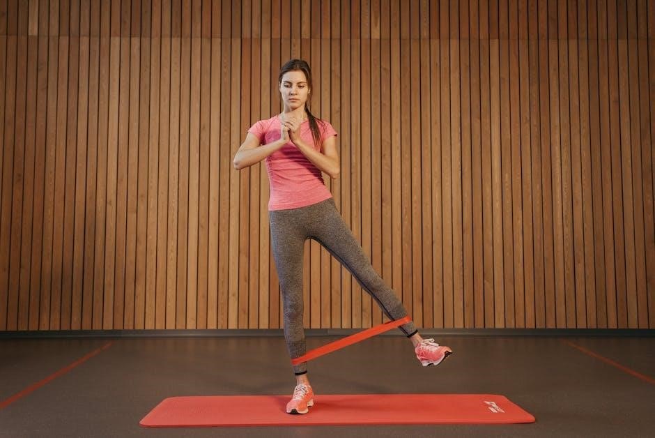
Hip Abduction and Adduction Exercises
Hip abduction and adduction exercises are vital for restoring balanced hip strength and stability, particularly after a labral tear. These movements target the muscles responsible for moving the leg away from (abduction) and towards (adduction) the midline of the body.
Initially, these exercises are performed with resistance bands, focusing on controlled movements and avoiding pain. Side-lying leg raises (abduction) and standing hip adduction with a band are common starting points.
Progression involves increasing band resistance, adding repetitions, and incorporating exercises like clam shells. Maintaining proper form—avoiding hip hiking or rotation—is crucial. Strengthening these muscles supports the hip joint, improves functional movement, and contributes to long-term stability.
Gluteal Muscle Strengthening
Gluteal muscle strengthening is paramount in hip labral tear rehabilitation, as these muscles provide crucial support and stability to the hip joint. Weak glutes contribute to poor biomechanics and can exacerbate impingement.
Exercises begin with foundational movements like glute bridges, progressing to single-leg variations for increased challenge. Hip thrusts, utilizing bodyweight or added resistance, effectively target the gluteus maximus.
Lateral band walks and clam shells focus on the gluteus medius, essential for hip abduction and pelvic stabilization. Proper form—engaging the glutes and maintaining a neutral spine—is vital. Strong glutes improve hip control, reduce stress on the labrum, and facilitate a return to functional activities.
Phase 4: Return to Activity (16+ Weeks)
Sport-specific training and agility drills are introduced, with a gradual increase in activity, ensuring the hip can withstand demands post-rehabilitation.
Sport-Specific Training
Sport-specific training marks a crucial phase, bridging rehabilitation and full athletic return. This stage meticulously replicates the movements and demands of the individual’s chosen activity, progressively challenging the repaired hip joint. For runners, this involves interval training, gradually increasing distance and pace. Soccer players will focus on cutting, pivoting, and shooting drills.
The intensity and complexity of these drills are carefully monitored, guided by pain levels and functional assessments. Emphasis remains on maintaining proper biomechanics to avoid re-injury. Exercises should mimic game situations, building confidence and neuromuscular control. A physical therapist designs a tailored program, ensuring a safe and effective transition back to the athlete’s specific sport, considering the nuances of their position and playing style.
Agility Drills
Agility drills are paramount in restoring dynamic hip control and preparing for the unpredictable demands of daily life and athletic pursuits. These exercises progressively challenge the hip’s ability to rapidly change direction, accelerate, and decelerate. Initial drills might include cone drills, focusing on lateral movements and figure-eight patterns.
As strength and proprioception improve, more complex drills are introduced, such as shuttle runs and reactive agility exercises. These drills require quick decision-making and coordinated movements. Proper form is crucial to prevent compensatory patterns and minimize stress on the healing labrum. A physical therapist guides progression, ensuring drills are performed safely and effectively, building confidence and functional capacity.
Gradual Increase in Activity Level
Returning to activity following hip labral tear rehabilitation demands a meticulously planned, gradual progression. Avoid sudden increases in intensity or duration, as this can re-irritate the healing tissues. Begin with low-impact activities like walking, cycling, or swimming, monitoring for any pain or discomfort.
Incrementally increase the duration and intensity of these activities over weeks, guided by symptom response. Reintroduce sport-specific movements slowly, focusing on proper technique. Listen to your body and respect pain signals; setbacks are common, and modifying the program is essential. A physical therapist provides personalized guidance, ensuring a safe and sustainable return to desired activity levels.
Phase 5: Maintenance and Prevention
Long-term success relies on a consistent exercise program strengthening core and hip muscles, alongside understanding hip anatomy to prevent future labral injuries.
Long-Term Exercise Program
Maintaining hip health post-rehabilitation requires a dedicated, ongoing exercise program. This isn’t about quick fixes, but building sustainable strength and stability. Focus should remain on core stabilization, incorporating exercises like planks, bridges, and side-lying hip abductions.
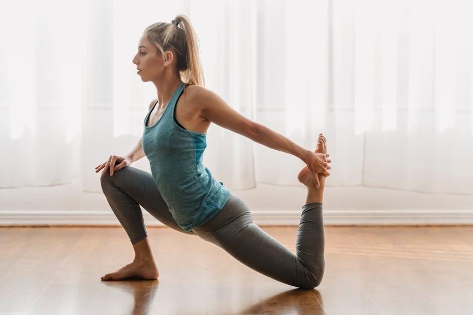
Continue progressive strengthening, including squats, lunges, and deadlifts – performed with proper form to avoid re-injury. Regular stretching, targeting hip flexors, hamstrings, and glutes, is crucial for maintaining range of motion.
Proprioceptive exercises, like single-leg balance drills, enhance joint awareness and control. A consistent routine, 2-3 times weekly, will help prevent future labral tears and maintain optimal hip function; Remember, listen to your body and modify exercises as needed;
Hip Joint Anatomy Review
Understanding the hip’s structure is key to comprehending labral tears. The hip joint is a ball-and-socket joint formed by the femoral head – the “ball” at the top of the thigh bone (femur) – and the acetabulum, a socket within the pelvis.
The pelvis itself comprises three fused bones: the ilium, pubis (pubic bone), and ischium. These converge to create the acetabulum, providing a stable base for the femoral head. The labrum, a ring of cartilage, deepens the acetabulum, enhancing stability and shock absorption.
Tears often occur where the femoral head impacts the acetabulum, particularly with FAI. Knowing these components helps visualize how injuries happen and why specific exercises target these areas for healing and prevention.
Understanding Femoroacetabular Impingement (FAI)
Femoroacetabular Impingement (FAI) is a frequent contributor to hip labral tears. It occurs when there’s abnormal contact between the femoral head and the acetabulum during hip movement. This abnormal contact can damage the labrum, leading to pain and limited range of motion.
There are three main types of FAI: cam, pincer, and mixed. Cam impingement involves an abnormally shaped femoral head, while pincer impingement relates to an overcovered acetabulum. Mixed FAI combines both.
FAI often develops gradually, and early intervention is crucial. Understanding the specific type of FAI is essential for tailoring rehabilitation programs, including targeted exercises to improve hip mechanics and reduce impingement.
Post-Operative Considerations
Post-operative rehabilitation following hip arthroscopy for a labral tear is a phased process, typically spanning several months. The success of surgery heavily relies on adherence to a structured protocol guided by a physical therapist.
Early phases (0-6 weeks) focus on pain and swelling management, gentle range of motion, and isometric exercises. Progression is gradual, advancing to strengthening and proprioceptive training (6-12 weeks).
Factors influencing recovery include whether the labrum was repaired or reconstructed. A comprehensive 5-phase program, as outlined in patient-guided protocols, is often employed. Returning to sport-specific activities requires careful assessment and a gradual increase in activity levels.
Importance of Patient Guidance
Effective rehabilitation hinges on strong patient engagement and understanding of the protocol. A patient-guided program, like the comprehensive 5-phase approach, empowers individuals to actively participate in their recovery.
Clear communication between the patient, surgeon, and physical therapist is crucial. Modifications to the exercise regimen should be based on individual progress and pain levels, always under professional discretion.
Adherence to the prescribed timeline and exercise intensity is paramount. Consistent effort, coupled with diligent self-monitoring, maximizes the potential for successful outcomes. Patient education regarding hip anatomy and FAI contributes to informed decision-making throughout the rehabilitation journey.
The Role of Physical Therapy
Physical therapy (PT) is central to both prehabilitation and post-operative recovery following hip arthroscopy for labral tears and FAI. A structured PT protocol, often spanning five phases, guides patients through progressive exercises.
PT interventions focus on restoring range of motion, strengthening hip and core muscles, and improving proprioception – the body’s awareness of its position in space. Therapeutic exercises are tailored to individual needs and progress.
PTs provide crucial guidance on proper form, pain management, and activity modification. They monitor patient response and adjust the program accordingly, ensuring a safe and effective rehabilitation process.
Hip Bone Structure (Ilium, Pubis, Ischium)
The hip bone, also known as the coxal or innominate bone, is a large, irregular structure forming the pelvis. It’s comprised of three fused bones: the ilium, pubis, and ischium. These bones converge to create the acetabulum, a deep socket crucial for hip joint stability.
The ilium forms the upper, flared portion of the hip bone, while the pubis constitutes the lower, anterior part. The ischium makes up the lower, posterior section, including the “sit bones.”
Understanding this structure is vital, as the acetabulum’s shape and alignment influence hip mechanics and can contribute to conditions like femoroacetabular impingement (FAI), often linked to labral tears.
Femoral Head and Acetabulum
The hip joint is a ball-and-socket joint formed by the femoral head – a ball-shaped bone at the femur’s top – and the acetabulum, a socket within the pelvis. This articulation allows for a wide range of motion, essential for daily activities.
The femoral head’s smooth cartilage covering enables frictionless movement within the acetabulum. The labrum, a ring of cartilage, deepens the acetabulum, enhancing stability and shock absorption.
Improper alignment or shape of either the femoral head or acetabulum can lead to femoroacetabular impingement (FAI), a common cause of hip labral tears. Understanding this relationship is key to targeted rehabilitation.